What Is a Hoof Abscess?
The biology behind one of the most painful — and most misunderstood — hoof conditions.
🔬 Why Abscesses Hurt So Much
A hoof abscess is a localized pocket of pus — bacteria, dead white blood cells, and cellular debris — trapped inside the hoof capsule. Unlike a skin abscess on the body, which can swell outward, the abscess inside a hoof is surrounded on all sides by rigid, unyielding horn. As bacteria multiply and the body's immune response floods the area with fluid, pressure inside the pocket builds rapidly. That pressure — with nowhere to expand — compresses the sensitive laminae, corium, and digital cushion against hard hoof wall. The result is intense, acute pain completely disproportionate to the actual amount of infection involved.
The abscess typically forms in one of two locations: subsolar (beneath the sole, between the sole and the sensitive corium) or submural (beneath the hoof wall, between the wall and the sensitive laminae). Both are equally painful; submural abscesses tend to track upward and break out at the coronary band if not drained from below.
Abscesses are self-limiting — left untreated, the body will eventually force the abscess to break out on its own, usually at the coronary band (the "gravel" that old-timers called it). Drainage relieves the pressure immediately and dramatically. A horse that was three-legged lame in the morning is often trotting sound by afternoon after the abscess is opened.
🦠 The Bacteria Involved
The most common organisms are Fusobacterium necrophorum and Bacteroides species — the same anaerobic bacteria responsible for thrush. They enter through a break in the white line, a nail hole, a bruise, or any compromise in the sole or wall that creates an anaerobic environment. Once established, they multiply rapidly in the oxygen-free space between layers of hoof tissue, producing gas and pus that builds under pressure.
📍 Where They Form
Abscesses most commonly form:
- White line zone — the most common entry point; soft, porous white line tissue allows bacteria to track inward from the ground surface
- Beneath old nail holes — pulled shoes leave nail holes that can trap debris and bacteria
- Beneath bruised sole — a subsolar bruise from a stone or hard impact creates damaged tissue where bacteria can establish
- Seat of corn — between the bar and the hoof wall at the heel; corns from poorly fitted shoes are frequent abscess sites
- Coronary band tracking — a deeply established abscess works upward through the hoof, breaking out at the coronary band
🎯 Recognizing an Abscess
Classic abscess presentation:
- Sudden onset — sound yesterday, severely lame today; the overnight change is a hallmark
- Non-weight-bearing or toe-touching only — the horse refuses to load the heel where pressure is worst
- Bounding digital pulse — detectable pulse at the back of the fetlock; stronger than normal, often strong enough to feel without pressing hard
- Hot hoof — the affected foot is noticeably warmer than the opposite foot
- Positive hoof tester response — the farrier or vet applies hoof testers across the sole; the horse flinches sharply when pressure hits the abscess location
- Swelling above the hoof — in some cases, a tracking abscess causes fetlock or pastern swelling as it works upward
What Causes Hoof Abscesses
Abscesses have specific, identifiable triggers. Understanding the cause helps prevent the next one.
Wet-Then-Dry Ground Conditions
The most common seasonal trigger. When the ground goes from very wet (winter rain, snow melt) to dry and hard (spring or summer drought), the white line softens in wet conditions and then hardens around trapped bacteria as the ground dries. The rapid change in ground hardness also drives grit and bacteria deeper into existing white line separations. Spring and autumn are peak abscess seasons for this reason.
Nail Pricks and Close Nails
A nail driven into or very close to the sensitive laminae during shoeing — a "prick" or "close nail" — introduces bacteria directly into sensitive tissue. The farrier may not realize a nail was placed too close until the horse goes lame 24–72 hours later. A nail prick abscess tends to be severe and rapid in onset. When a freshly shod horse goes three-legged lame within days, the shoe comes off first and nail placement is investigated.
Stone Bruise
A direct impact to the sole from a sharp stone, gravel, or hard ground surface creates a subsolar bruise — damaged, hemorrhagic tissue beneath the sole. Bruised tissue is more susceptible to bacterial colonization. The bruise itself causes immediate pain, but an abscess may develop days to weeks later as bacteria establish in the damaged tissue. Horses with thin soles, flat soles, or those worked on rocky terrain are highest risk.
Laminitis Complication
Chronic laminitis creates a stretched, compromised white line and unstable lamellar junction — a perfect entry point for bacteria. Laminitic horses have significantly higher abscess rates than healthy horses, often developing multiple abscesses in the same feet repeatedly. In a horse with a history of laminitis, recurrent abscesses are almost always related to the ongoing white line compromise.
Poorly Fitted or Overdue Shoe
A shoe that has shifted, spread, or grown past the hoof wall creates pressure points. The seat of corn — the angle between the bar and hoof wall at the heel — is particularly vulnerable to pressure from a shifted shoe or an overlong shoeing interval where the heel grows over the shoe. Corns at the seat of corn are a frequent abscess site in shod horses.
Foreign Body Penetration
A sharp object penetrating the sole — a nail, wire, thorn, or sharp stone that the horse steps on — drives bacteria deep into hoof tissue. Sole penetrations are more serious than typical abscesses because the wound tract can reach deeper structures including the coffin joint, navicular bursa, or digital flexor tendon sheath. Any puncture wound to the sole is a veterinary emergency until the depth and direction are confirmed.
White Line Disease Complication
Established white line disease creates hollow, separated cavities in the hoof wall — pockets that can fill with debris and bacteria and become abscesses. A horse with chronic WLD that suddenly goes lame often has a WLD-related abscess at the site of existing separation. Treatment requires addressing both the abscess and the underlying WLD.
Hoof Imbalance
A chronically imbalanced foot — with one heel consistently higher or one quarter bearing more load — creates micro-trauma and bruising in the overloaded areas. Over time, this repetitive low-grade injury creates tissue damage that bacteria can colonize. Horses with persistent medial-lateral imbalance often develop recurrent abscesses in the same overloaded quarter.
Immunosuppression
Horses with PPID (Cushing's disease) or severe systemic illness have reduced immune function — they are less able to contain minor bacterial contamination before it escalates to a full abscess. PPID horses develop hoof abscesses at higher rates than healthy horses, often in conjunction with the chronic laminitis and white line compromise that PPID causes. This is another reason PPID management with pergolide matters.
⚡ Quick Reference — Hoof Abscess at a Glance
Treatment — Three Roles, Three Approaches
Drainage is the cure for an abscess. The farrier or vet provides it; the owner maintains the aftercare that keeps the drainage site clean and the abscess from reforming.
The Farrier's Role — Finding and Draining the Abscess
For a typical hoof abscess with no sole penetration history, the farrier is the first call — not the vet. Experienced farriers locate and drain abscesses routinely. The entire procedure, once the abscess is located, takes minutes and provides immediate dramatic relief.
If the horse is shod, the shoe comes off first. This allows full access to the sole and white line for examination, and removes any shoe pressure that may be compressing the abscess site. Shoe removal alone sometimes provides noticeable relief if the abscess is at the seat of corn or a pressure point beneath the shoe branch.
The farrier applies hoof testers — a large pincer instrument — methodically across the sole, pressing each section in turn. The horse reacts sharply (flinching, pulling the foot away, or showing visible discomfort) when pressure is applied over the abscess. The farrier narrows the location to a specific quadrant of the sole, then examines the area visually:
- A dark, discolored spot in the white line or sole — often appearing as a gray or black tract
- A soft or "punky" feel when the sole is pressed with a hoof knife
- A visible tract or tunnel in the white line leading inward
- In some cases, pus or a dark fluid is already visibly tracking through the white line
Once located, the farrier uses a hoof knife to carefully follow the tract inward, paring away overlying sole or white line horn until the abscess cavity is reached. The goal is to create a drainage hole large enough to allow complete emptying of the pus pocket — typically the diameter of a pencil or slightly larger. When the abscess is breached, pus (a black, dark gray, or blood-tinged fluid) drains out under pressure. The horse's relief is often visible immediately — the tension in the leg drops, and the horse may begin to bear weight within minutes of drainage.
Key principle: open it wide enough. A drainage hole that is too small seals over with new horn quickly, trapping residual infection. A properly sized opening allows full drainage and remains open long enough for the cavity to close from the inside out.
After drainage, the farrier flushes the abscess cavity with a dilute antiseptic solution — typically 2% chlorhexidine, dilute povidone-iodine (Betadine), or hydrogen peroxide — using a syringe to reach into the cavity. This removes residual pus and kills remaining bacteria. Flushing is repeated at the first owner soaking session and at subsequent farrier visits.
The drained cavity is packed with an antiseptic drawing agent — ichthammol (black salve), povidone-iodine soaked cotton, or a commercial poultice — to continue drawing out any residual infection and keep the drainage site clean. The foot is then wrapped to protect the sole and keep the packing in place:
- Cotton or gauze over the packed cavity
- Duct tape or hoof boot over the sole to protect from ground contact
- Bandage wrap around the hoof and coronary band to hold everything in place
- The farrier instructs the owner on the soaking and rewrapping protocol for home care
Not every abscess is immediately findable. If hoof testers are positive but no visible tract appears after careful examination, the farrier may:
- Recommend 24–48 hours of soaking to soften the sole and draw the abscess closer to the surface
- Apply a poultice wrap overnight to "ripen" the abscess
- Refer to a vet for nerve block and more intensive examination if the horse is not improving after 24–48 hours of treatment
Hoof Testers
Large pincer tool applied across the sole to localize abscess pain. The primary diagnostic tool for abscess location. Every farrier carries them; owners can learn to use a simplified version (thumb pressure) but hoof testers are far more precise.
Ichthammol (Black Drawing Salve)
Sulfur-based drawing agent packed into the abscess cavity after drainage. Pulls residual infection toward the surface, softens surrounding horn, and has mild antiseptic properties. Applied with cotton packing and held in place with a wrap.
Animalintex Poultice Pad
Commercial ready-to-use poultice pad. Moistened and applied to the sole after drainage — draws infection, softens horn, and maintains warmth at the abscess site. Standard recommendation for owner home-care soaking and wrapping.
Epsom Salt (Magnesium Sulfate)
Dissolved in warm water for daily hoof soaking. Draws out infection through osmotic action, softens the sole to facilitate drainage, and reduces pain. 1–2 cups per gallon of comfortably warm water, soak 15–20 minutes twice daily.
The Veterinarian's Role — When an Abscess Becomes More Than an Abscess
Most hoof abscesses do not require a vet. The vet becomes essential when the abscess involves a sole puncture wound, when it fails to resolve with farrier treatment, when swelling is spreading up the leg, when the horse has a fever, or when deep structures may be involved.
A horse that has stepped on a nail, wire, or other sharp object penetrating the sole is a veterinary emergency — not a farrier call first. The critical issue is the trajectory of penetration:
- A puncture in the front two-thirds of the sole risks penetrating the coffin joint
- A puncture in the middle third risks the navicular bursa
- A puncture near the frog risks the digital flexor tendon sheath
Infection in any of these synovial structures (joint, bursa, tendon sheath) is a surgical emergency with a guarded to poor prognosis. If a nail or object is still in the foot: do not remove it — call the vet immediately and keep the horse still. The vet will take radiographs with the object in place to determine the direction and depth of penetration before removal. Removing the object first destroys the most important diagnostic information.
When a farrier cannot locate the abscess despite positive hoof tester response, the vet can administer a palmar digital nerve block to desensitize the foot — confirming that the pain source is inside the hoof — and then assist with more aggressive sole examination. Radiographs can sometimes reveal gas pockets inside the hoof that indicate abscess location not visible from the sole surface. In rare cases, ultrasound can localize a soft-tissue abscess tracking up the pastern.
Routine hoof abscesses do not require systemic antibiotics — drainage is the cure, and antibiotics cannot reach an avascular abscess cavity. However, antibiotics are appropriate when:
- The abscess has penetrated a synovial structure (joint, bursa, tendon sheath)
- There is cellulitis — spreading infection up the lower limb with heat, swelling, and pitting edema
- The horse has a fever (rectal temperature above 38.5°C / 101.5°F)
- The horse is immunocompromised (PPID) and the infection is not resolving normally
For abscesses that have penetrated synovial structures, regional limb perfusion — injecting high-concentration antibiotic directly into the blood vessels of the lower limb with a tourniquet applied above — delivers antibiotic concentrations to infected tissue that systemic dosing cannot achieve. This technique is used in hospital settings for serious foot infections and is often combined with surgical lavage (flushing) of the infected joint or tendon sheath.
For horses with severe abscess pain who are not improving within 24 hours of drainage, or for horses with spreading cellulitis, the vet may prescribe:
- Phenylbutazone (bute) — 1–2 grams twice daily for 3–5 days; reduces pain and systemic inflammation
- Flunixin meglumine (Banamine) — particularly for horses with fever or systemic signs
If your horse has a nail or sharp object in its foot, the natural instinct is to remove it immediately. Resist this. Radiographs taken with the object in place show exactly where it went — information that determines whether the horse needs emergency surgery or can be treated conservatively. Removing the nail first destroys that information and can make surgery more complicated. Call your vet, keep the horse still and calm, and wait for professional guidance. If you cannot reach a vet and the horse is in extreme distress, photograph the nail in situ from multiple angles before removal.
The Horse Owner's Role — Soaking, Wrapping, and Aftercare
Once the farrier has drained the abscess, the owner takes over daily aftercare — soaking and rewrapping to keep the drainage site open, clean, and drawing out residual infection. This is genuinely effective work that dramatically speeds recovery. The better the owner's aftercare, the faster the horse returns to soundness.
Soaking is the cornerstone of abscess aftercare. Here is the correct protocol:
- Fill a rubber feed tub, soaking boot, or bucket with comfortably warm water — warm enough that you can hold your hand in it comfortably
- Dissolve 1–2 cups of Epsom salt (magnesium sulfate) per gallon of water — this creates an osmotic solution that draws infection from tissue
- Stand the horse in the bucket for 15–20 minutes, twice daily
- Keep the water warm — add hot water as it cools; cold water soaking is ineffective and uncomfortable
- After each soak, dry the hoof thoroughly and apply fresh packing and wrapping before returning the horse to its stall or paddock
- Continue soaking for 2–3 days after the horse appears fully sound — the abscess cavity needs time to close from the inside out
Between soaking sessions, a poultice wrap continues the drawing action and protects the drainage site. Step-by-step:
- After the soak, dry the hoof thoroughly — wet wrapping traps moisture and causes softening
- Apply ichthammol (black salve) or a wet Animalintex pad directly over the drainage site — these drawing agents pull residual infection toward the surface
- Cover the sole with cotton or gauze to hold the poultice in place
- Wrap the entire hoof with Vetwrap or Elastikon, going up onto the pastern for security
- Apply duct tape in an X pattern across the bottom of the wrap to protect it from the ground — without duct tape, the wrap lasts minutes in a stall
- Check and replace the wrap twice daily when soaking; check daily once the horse is more comfortable and soaking has ended
An open abscess drainage site is a wound — it needs a clean environment:
- Keep the horse in a dry, clean stall with fresh bedding, not in a muddy paddock where the drainage site can become contaminated
- Change bedding daily during active treatment
- If turnout is necessary, use a hoof boot over the wrap to protect it from mud and debris, and limit the turnout time
- Do not ride until the horse is fully comfortable without the wrap and has been sound for at least 48 hours
Normal recovery progression:
- Day 1 after drainage: Significant improvement — horse bearing weight, digital pulse settling
- Days 2–3: Progressively more comfortable; walking normally; digital pulse nearly normal
- Days 4–7: Sound or nearly sound; drainage site closing; wrap still protective
- 1–3 weeks: Full soundness; drainage site fully closed; back to normal management
Call your farrier or vet if: the horse is not improving after 48 hours of soaking, the lameness is getting worse rather than better, you see swelling spreading above the hoof, the horse develops a fever, or a second abscess appears to be forming in the same foot.
Epsom Salt
Magnesium sulfate. 1–2 cups dissolved in warm water per gallon. The osmotic action draws infection from tissue. Inexpensive, widely available, genuinely effective. The most important owner tool for abscess aftercare.
Ichthammol (Black Drawing Salve)
Sulfur-based drawing agent. Applied directly to the drainage site between soaks. Pulls residual infection toward the surface and maintains antiseptic contact with the healing cavity. Available at farm supply stores.
Animalintex Poultice Pad
Ready-to-use kaolin poultice pad. Moistened with warm water and applied to the sole. Draws infection, maintains warmth, and conforms to the sole surface. Easier to use than mixing your own poultice. Replace daily.
Hoof Soaking Boot
A rubber or EVA boot that seals around the pastern, allowing the hoof to be soaked without a bucket. The horse can stand relatively free during the soak. Much easier for one person to manage than a bucket, especially with a moving or impatient horse.
Abscesses strike without warning, always at an inconvenient hour. Keep an abscess kit stocked and ready: Epsom salt (a full bag), ichthammol, Animalintex pads, cotton or gauze squares, Vetwrap, duct tape, and a rubber soaking tub or boot. When your horse goes three-legged lame at 6pm on a Sunday, having everything on hand means you can start soaking immediately while you wait for your farrier to call back — and that matters.
Prevention — Reducing Abscess Risk
No management eliminates abscess risk entirely, but consistent practices dramatically reduce frequency — especially in horses that are prone to recurrence.
Daily Hoof Picking
The most effective single prevention. Daily picking removes the packed debris, grit, and manure that drives bacteria into white line separations. A two-minute daily routine prevents weeks of abscess recovery. This is the baseline — no other prevention measure replaces it.
Dry, Clean Living Environment
Chronic wet conditions soften the white line and create the anaerobic pockets that bacteria colonize. Deep, dry bedding; clean stalls; gravel footing in high-traffic paddock areas. Eliminating prolonged standing in mud and wet manure is the most impactful environmental change for abscess-prone horses.
Regular Farrier Schedule
Regular trimming maintains the white line integrity and sole thickness that resist bacterial penetration. An overlong hoof with a distorted, flared wall and weak white line is far more abscess-prone than a well-maintained foot. 6–8 week maximum interval; 5–6 weeks for horses with abscess history.
Correct Hoof Balance
Medial-lateral imbalance creates repetitive bruising in overloaded quarters — a recurring abscess trigger. Ask your farrier to specifically assess and correct balance at every visit, not just length. A balanced foot distributes load evenly and reduces the micro-trauma that bacteria exploit.
Manage Underlying Conditions
Horses with laminitis, WLD, or PPID have chronically compromised hoof defenses. Managing these underlying conditions — dietary control for laminitis, Prascend for PPID, antifungal treatment for WLD — directly reduces abscess frequency in affected horses.
Protective Footing and Sole Pads
Horses with thin soles working on rocky terrain benefit from full sole pads and pour-in packing — protecting the sole from the stone bruises that initiate subsolar abscesses. For trail horses or horses regularly on gravel, this is standard preventive care, not an optional extra.
Hoof Abscess Videos — Watch & Learn
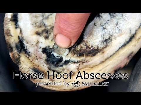
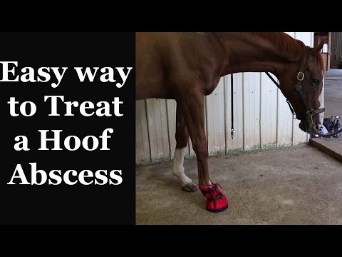
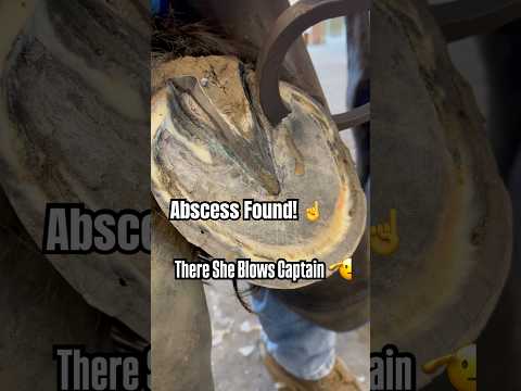
See abscesses located, drained, and wrapped — and learn the home soaking technique from experienced horse owners and farriers.