What shoes are used, why they work, what to expect at each appointment, and what realistic outcomes look like — for every major therapeutic shoeing condition.
What Therapeutic Shoeing Is
Therapeutic shoeing is the deliberate use of shoe design, material, pads, wedges, and modifications to manage or treat a lameness condition. It is not a standalone treatment — it is part of a coordinated plan between the veterinarian who diagnoses the condition and the farrier who executes the mechanical response to that diagnosis.
The shoe becomes a tool of precision medicine. Applied correctly with imaging guidance and veterinary oversight, therapeutic shoeing can reduce pain dramatically, slow or halt structural damage, support tissues through healing, and in many cases allow horses to return to useful work that would otherwise be impossible. Applied incorrectly — without diagnosis, without imaging, without vet-farrier communication — it can mask symptoms, delay proper treatment, and cause harm.
This guide covers the seven most common therapeutic shoeing conditions — what the farrier does, what shoes are used, what each shoe achieves, what owners experience during the process, and what outcomes are realistic. For the underlying philosophy and five mechanical variables that all therapeutic shoeing manipulates, see the Corrective & Therapeutic Shoeing Overview →
The Non-Negotiable Foundation
Every therapeutic shoeing plan in this guide assumes a veterinary diagnosis has been made, imaging (radiographs at minimum) has been obtained, and the farrier and vet are in direct communication. Therapeutic shoeing without these foundations is guesswork. It may accidentally help — or it may cause harm while the actual problem progresses.
What Happens at a Therapeutic Appointment
A therapeutic shoeing appointment is meaningfully different from a routine visit. Owners who understand what to expect can prepare properly, observe usefully, and communicate productively.
1
Movement Assessment
The farrier watches the horse walk and often trot before touching a foot. In a therapeutic case, any change in gait quality since the last visit is meaningful data — it tells the farrier whether the current shoeing is working. Note what you observe between appointments and report it accurately.
2
Hoof Assessment
The farrier examines the hoof carefully before any trimming — hoof wall quality, heel condition, frog health, sole depth (using hoof testers if indicated), wall temperature, coronary band condition, and how the current shoe has worn. Changes since the last visit are noted and factored into the plan for this cycle.
3
Radiograph Review (if applicable)
For laminitis, navicular, and any condition where the coffin bone position matters, the farrier should review current radiographs before trimming. In an ongoing case, this may be a review of films taken that visit by the vet; in a stable managed case, the films from the last veterinary visit. The trim should be guided by what the imaging shows, not only by what the external hoof looks like.
4
Trimming to Therapeutic Parameters
The trim in a therapeutic case is calculated — specific amounts removed from specific locations, often measured against the radiograph. For a laminitic horse, the trim may include dorsal wall removal (resection) to eliminate leverage on rotating laminae. For a navicular horse, careful heel management to prevent collapse. This takes longer than a routine trim and requires more explanation — ask questions.
5
Shoe Preparation
Therapeutic shoes may need to be modified — bars welded, toes squared or rolled, wedge pads fitted, clips added, or in complex cases, shoes custom-forged from bar stock entirely. A therapeutic appointment can take 90 minutes to two hours per horse for complex cases. The time is not inefficiency — it is precision.
6
Application & Check
After shoeing, the farrier checks the fit carefully and usually watches the horse move again. For laminitis cases, post-shoeing comfort assessment is critical — a horse that was comfortable before the farrier and sore after indicates something needs adjusting before the farrier leaves.
7
Debrief and Next Steps
A good therapeutic farrier will tell you what they did, why, what to watch for, and when to call if something changes. They will communicate with your vet about what they observed. The next appointment should be scheduled before the farrier leaves — therapeutic horses cannot run past their interval.
Laminitis & Founder
Laminitis & Founder
Inflammatory failure of the lamellar bond — coffin bone rotation and sinking
⚕️ Therapeutic
Laminitis is the most complex and consequential therapeutic shoeing situation a farrier faces. The inflammatory failure of the lamellae — the interlocking tissue that suspends the coffin bone inside the hoof capsule — allows the coffin bone to rotate under the pull of the deep digital flexor tendon, or to sink toward the ground surface, or both. The shoeing objectives are to relieve pressure on the rotating toe laminae, support the sinking coffin bone, and reduce the DDFT tension that drives further rotation.
Every laminitis shoeing plan must be guided by radiographs. The rotation angle, sole depth remaining, and coffin bone position determine what interventions are possible and in what order. A farrier working on a laminitic horse without radiographs is making critical decisions blind.
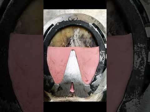
Heart Bar Shoe
Loads the frog to transfer weight from the compromised lamellar zone to the frog and digital cushion. The bar must contact the frog with calibrated pressure — calculated from sole depth on radiograph. Too much pressure causes frog pain; too little provides no benefit. Requires precise fitting and a farrier experienced with laminitis.
Reverse Shoe
Applied toe-heel reversed — the branches of the shoe extend behind the foot, eliminating all leverage at the toe while providing rear support. Counterintuitive but highly effective for moderate to severe rotation. Eliminates the toe breakover entirely.
Wedge Pad / Elevated Heel
Reduces DDFT tension by reducing the angle differential between the DDFT and the ground. The degree of elevation is calculated from the rotation angle on radiograph. Typically 4–10 degrees. Combined with a rolled or squared toe to ease breakover from the elevated position.
Pour-In Pad
Polyurethane material injected under the shoe and allowed to set against the sole, providing uniform sole support and protecting the dropping coffin bone from ground contact. Particularly valuable as sole depth decreases in severe rotation or sinking cases.
Glue-On Shoe System
When hoof wall quality fails — common consequence of repeated or severe laminitis — nailing is no longer viable. Glue-on composite shoes bond to the remaining sound wall, distribute load through the cuff, and eliminate nail-hole stress in fragile wall. The Imprint and Dalric systems are the most widely used in laminitis management.
Dorsal Wall Resection
Removing the outer hoof wall at the toe eliminates the mechanical leverage pulling the laminae apart as the coffin bone rotates. Combined with a protective dressing and pad while new wall grows in from above. Not a shoe type — a surgical trimming intervention that fundamentally changes what shoe can then be applied.
Realistic outcome: Management, not cure. Horses with mild to moderate rotation that receive prompt, correct treatment can return to full work. Severe rotation or sinking carries a guarded prognosis. Plan in months to years, not weeks. Repeated radiographs every 2–3 shoeing cycles track whether the coffin bone is stabilizing.
For the full clinical guide including acute phase management and long-term protocols, see Laminitis & Founder →
Navicular Syndrome
Navicular Syndrome
Palmar foot pain — the leading cause of chronic front-end lameness
⚕️ Therapeutic
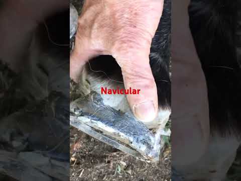
Navicular syndrome encompasses pain originating in the navicular bone, navicular bursa, deep digital flexor tendon at its insertion, and the associated soft tissue structures of the palmar foot. The common thread is that these structures are loaded and stressed at every footfall — and in navicular horses, that loading produces pain. Therapeutic shoeing cannot reverse the bony changes that characterize advanced navicular disease, but it can significantly reduce the mechanical stress that causes pain at each stride, making many navicular horses comfortable and usefully working for years.
The three mechanical targets: reduce DDFT tension (raise the heel), extend the base of support palmarly (egg bar), ease breakover (rolled or squared toe). Most navicular protocols address all three simultaneously.
Egg Bar Shoe
The most important single modification for navicular syndrome. The extended oval bar behind the heels moves the base of support rearward, reducing the load on the navicular apparatus and providing a more stable landing surface. Many navicular horses show dramatic comfort improvement after the first egg bar application. Requires a competent farrier — a poorly fitted egg bar that extends too far can lever the shoe off; one that doesn't extend enough provides little benefit.
Wedge Pad (2–6 degrees)
Raises the heel angle, reducing the angle of the DDFT as it passes over the navicular bone and reducing compressive load on the navicular bursa. Used with egg bars in most navicular protocols. The degree of elevation is a clinical judgment — too much can cause heel pain, too little provides inadequate relief.
Rolled or Rockered Toe
Moving the breakover point toward the heel reduces the peak DDFT loading at push-off — the phase of stride when DDFT tension is highest. Simple to achieve on any shoe type; highly effective for reducing palmar foot pain. Often the modification that produces the most immediate visible comfort improvement.
Wide Web Shoe
Distributes ground contact load across a wider bearing surface, reducing peak pressure at any one point on the hoof wall. Useful when the palmar foot is pressure-sensitive. Often combined with a full pad for horses with additional sole sensitivity.
Pour-In or Full Pad
Provides sole protection and cushioning. In navicular cases, the primary value is reducing ground impact force transmitted to the already-painful palmar structures at each footfall. Pour-in materials (polyurethane) provide more uniform sole contact than traditional leather or rubber pads.
Realistic outcome: Long-term management. Most navicular horses maintained on appropriate shoeing, appropriate work load, and 5–6 week intervals remain comfortable and working for years. The condition is chronic and progressive — the shoeing manages pain and mechanical contributors but does not halt the underlying bony changes. Interval compliance is critical: a navicular horse that goes 10–12 weeks between shoeings will often deteriorate significantly.
Tendon and ligament injuries require shoeing that reduces mechanical load through the injured structure during the healing period. The specific modification depends entirely on which structure is injured — each has a different anatomical path, loading pattern, and mechanical sensitivity. Ultrasound imaging of the lesion guides the shoeing decision as directly as radiographs guide laminitis management.
Heel Elevation (DDFT injuries)
The DDFT is loaded in tension as the heel drops at impact and in compression as it passes over the navicular bone. Raising the heel with wedge pads (4–8 degrees typically) reduces both the angular load and the compressive force at the navicular interface. The appropriate elevation is guided by the location and severity of the DDFT lesion on ultrasound.
Egg Bar + Heel Elevation (DDFT and SDFT)
The combination most commonly used for serious tendon injuries — the egg bar extends the base of support and reduces DDFT load while the heel elevation further reduces DDFT tension. For SDFT (superficial digital flexor) bows, the heel elevation reduces the loading on the superficial flexor during the weight-bearing phase.
Rolled Toe (all tendon injuries)
Eases breakover and reduces the peak DDFT tension at the push-off phase of stride. A simple modification compatible with any shoe type, valuable for all rear-of-foot and flexor tendon conditions.
Raised Heel (suspensory injuries)
Suspensory ligament injuries are exacerbated by fetlock drop — the downward travel of the fetlock under weight-bearing loads the suspensory maximally. Raising the heel reduces fetlock drop and therefore reduces peak suspensory loading with each stride. A less intuitive but well-supported application of heel elevation.
Realistic outcome: Highly variable by structure and severity. Superficial SDFT injuries heal over 6–18 months with appropriate rest and shoeing; deep DDFT tears and core lesions carry a more guarded return-to-work prognosis. Suspensory injuries in sport horses can end careers at upper levels. Shoeing supports healing but cannot accelerate it — compliance with exercise restriction is as important as the shoe choice.
Quarter Cracks
Quarter Cracks
Vertical wall splits at the quarter — caused by medial-lateral imbalance
⚕️ Therapeutic
Quarter cracks are vertical fissures in the hoof wall at the quarter that flex open and closed with every stride, pinching sensitive tissue between the edges and causing pain, occasionally bleeding, and infection risk. They are almost always caused by underlying medial-lateral imbalance — one quarter is loaded excessively because the foot is not landing evenly. Fix the crack without fixing the balance, and the crack returns.
Bar Shoe (straight or heart bar)
Connects the heels rigidly, preventing them from spreading and contracting independently at each footfall. This eliminates or significantly reduces the flexing at the quarter that causes crack edges to move against each other. The most basic and universally applied quarter crack shoe.
Wire or Fiberglass Lacing
Stainless steel wire or fiberglass strands are bridged across the crack, mechanically immobilizing the edges. The crack is cleaned and debrided first; the lacing prevents any relative movement regardless of loading. Highly effective when done well — requires skill and the right materials.
Acrylic or Epoxy Fill
After the crack is stabilized and any infection cleared, the void can be filled with hoof acrylic to restore wall integrity and protect the sensitive tissue below. The fill is not structural — it doesn't prevent crack movement by itself — but combined with bar shoeing and lacing it maintains the repair while new wall grows in from above.
Quarter Clip
A clip positioned adjacent to the crack on the shoe prevents the hoof wall from sliding relative to the shoe during loading — a significant source of crack-flexing force. Quarter clips are a simple, effective addition to any quarter crack repair and are often the difference between a repair that holds and one that loosens.
Corrective Trimming to Equalize Balance
Not a shoe — but the most important intervention. Reducing the height of the overloaded quarter, equalizing medial-lateral balance, and addressing any flare in the affected wall corrects the cause. Without this, the crack returns after the current repair grows out.
Realistic outcome: Full resolution requires the crack to grow completely out from coronary band to ground surface — 9–12 months in most horses. The repair maintains soundness during this period. Recurrence is common if the underlying balance issue is not corrected and maintained.
Fungal/bacterial infection of the inner hoof wall — often requires significant resection
⚕️ Therapeutic
White line disease is a fungal and anaerobic bacterial infection of the non-pigmented inner layer of the hoof wall. Left untreated, it can excavate large areas of wall, leaving the foot with little structural integrity and no nail zone for conventional shoeing. Therapeutic shoeing for white line disease is largely about maintaining foot support when normal nailing is compromised or impossible.
Glue-On Shoe System
When the resected area eliminates the conventional nail zone, glue-on composite shoes bond to the remaining sound wall without nails. The cuff system distributes load across the available intact wall and provides the support the hoof needs while the resected area grows back from above. Most commonly used for toe or quarter resections.
Hospital Plate Shoe
A shoe with a removable plate that covers the sole — the plate can be removed at each treatment to access and medicate the resected area, then replaced to protect the foot between treatments. Allows ongoing wound management while maintaining foot support without re-shoeing at every treatment.
Fiberglass or Acrylic Reconstruction
Once the infection is completely eliminated and the cavity is verified clean, the void can be filled with fiberglass-reinforced acrylic to rebuild the missing wall structurally. This restores the nail zone (or enough of it for conventional shoeing) while the natural wall grows in from above. A well-executed reconstruction can be practically invisible and provides real structural support.
Realistic outcome: Good with prompt, thorough treatment. Small to moderate resections resolve fully with correct debridement, appropriate topical treatment, and shoeing to maintain support. Large toe resections take 12–18 months to fully resolve as the wall grows back. Recurrence is common in persistently wet, unhygienic environments — management change is as important as treatment.
Inadequate distance between coffin bone and ground — chronic bruising risk
⚕️ Therapeutic
Horses with thin soles have insufficient distance between the coffin bone and the ground surface of the sole. The consequence is chronic sole bruising on any hard or irregular surface, frequent abscesses as bruised tissue becomes infected, and in severe cases, direct coffin bone contact with the ground through a sole too thin to protect it. Therapeutic shoeing for thin-soled horses is about creating the barrier the sole cannot.
Full Pad (leather or synthetic)
A flat pad fitted between shoe and hoof wall covers the entire sole, preventing direct ground contact on any surface. Traditional leather pads compress over time and must be replaced at each shoeing. Modern synthetic materials (polyurethane, rubber composites) maintain their profile better and provide more consistent protection.
Pour-In Pad
Polyurethane compound injected after the shoe is applied, flowing into the sole and frog area and setting as a semi-rigid cushion. Provides more uniform sole contact than a flat pad, prevents debris packing under the shoe, and can be applied with or without a traditional pad depending on sole depth and sensitivity. The gold standard for severe thin soles.
Wide Web Shoe
A wider shoe branch covers more of the white line area, extending coverage slightly toward the sole and providing more bearing surface. Combined with pads in severe cases; used alone in milder situations where full sole coverage is not needed but peripheral sole protection is beneficial.
Realistic outcome: Management for most horses — sole depth is largely genetic and nutritional, and cannot be dramatically increased in horses with chronically flat or thin feet. Shoeing provides protection that makes the horse comfortable and working. Sole growth can be improved with correct nutrition (particularly adequate biotin and zinc) but the improvement is typically modest in horses with constitutional flat feet.
Bone proliferation and cartilage deterioration around lower limb joints
⚕️ Therapeutic
Ringbone — periosteal new bone production around the pastern or coffin joint — causes pain through joint capsule distension, abnormal mechanics during movement, and eventually cartilage destruction. Coffin joint arthritis without proliferative bone changes produces pain through the same mechanical pathway. Therapeutic shoeing targets the movement of the joint itself — reducing the range of motion stress at each stride reduces the pain produced.
Rolled Toe
The simplest and most immediately effective modification for coffin joint and low ringbone pain. The rolled toe allows the foot to roll forward at breakover rather than pivoting sharply, spreading the joint extension stress across a longer arc. Many horses show significant improvement from a rolled toe alone. Applicable to any shoe type as a modification.
Rocker Shoe (full rocker)
A shoe curved from heel to toe, allowing the foot to roll continuously in any direction with minimal forced joint extension. More aggressive than a rolled toe — used for moderate to advanced joint disease, bilateral joint involvement, or cases where a rolled toe alone is insufficient. The horse effectively stands on the apex of the rocker, with minimal point-loading anywhere in the stride cycle.
Wide Web with Full Pad
Reduces the concussive force transmitted to the affected joint at each footfall. Ground impact that reaches the joint is a significant pain contributor in arthritic horses. Pad materials vary in their shock-absorption properties — polyurethane and rubber absorb more than leather.
Realistic outcome: Depends heavily on location and severity. Low ringbone at the coffin joint has better prospects for comfortable long-term management than high ringbone at the pastern joint. Horses managed with rockers and joint injections commonly remain in comfortable light to moderate work for years. Coffin joint ankylosis (fusion) — when it occurs — can produce a surprisingly sound horse, as the fused joint no longer produces pain from motion.
Sheared Heels
Sheared Heels & Medial-Lateral Imbalance
One heel significantly higher — heels shear past each other under loading
⚕️ Corrective + Therapeutic
Sheared heels occur when one heel is significantly taller than the other, causing them to move independently under loading — shearing past each other rather than compressing together as designed. The consequence is intense focal stress at the higher quarter, driving quarter cracks, coronary band trauma, and palmar heel pain. The cause is almost always medial-lateral imbalance from trimming that has allowed one heel to grow taller than the other over multiple shoeing cycles.
Corrective Trimming to Equalize Heel Heights
The primary treatment — gradually reducing the taller heel toward the height of the lower heel over multiple shoeing cycles. Aggressive single-cycle correction causes lameness as the contracted structures adjust too rapidly. The farrier trims incrementally, aiming to equalize within 2–3 shoeing cycles.
Straight Bar Shoe
Connects both heels rigidly, preventing independent movement of the sheared heels regardless of landing pattern. Provides immediate mechanical relief while the trimming correction takes effect. The standard shoe for active sheared heel cases.
Quarter Clips (both sides)
Clips on both sides of the shoe prevent the hoof from sliding across the shoe surface during loading — a source of shearing force. Combined with bar shoeing, quarter clips on both sides provide comprehensive mechanical stability.
Wide Web at Affected Heel
A wider branch at the higher heel provides more bearing surface and distributes load more evenly at the painful quarter while the heel heights are being equalized.
Realistic outcome: Good with consistent management. Heel heights equalize progressively over 3–6 shoeing cycles when trimming is done correctly and the horse is maintained on a consistent interval. Without consistent trimming, sheared heels return — this is a condition that requires ongoing management, not a one-time fix.
Therapeutic Shoe Quick Reference
A summary of therapeutic shoe types and their primary applications.
Allows continuous rolling motion with minimal forced joint extension
Full Pad
Thin soles, bruising, impact sensitivity
Prevents direct sole-to-ground contact
Pour-In Pad
Laminitis, thin soles, navicular — uniform sole support
Distributes load uniformly across entire sole surface
Glue-On System
Laminitis (wall failure), white line resection, foals
Provides shoe support without nail-hole stress in compromised wall
Hospital Plate
White line disease, abscesses, severe wounds
Removable sole plate allows wound access while maintaining foot support
Wide Web
Thin soles, ringbone, concussion sensitivity
Distributes load over wider bearing surface, reduces peak pressures
Watch & Learn
Four videos covering therapeutic shoeing in practice — from laminitis management to egg bar application and corrective work. Tap any thumbnail to play.