What Is Navicular Syndrome?
The anatomy, the structures involved, and why "navicular disease" is now considered an outdated term.
🔬 The Navicular Apparatus — Four Structures, One Problem
The navicular bone (distal sesamoid bone) is a small, boat-shaped bone nestled at the back of the horse's foot, sitting behind the coffin joint. It acts as a pulley over which the deep digital flexor tendon (DDFT) passes on its way to attach to the bottom of the coffin bone. Cushioning this tendon-bone interface is the navicular bursa, a fluid-filled sac that reduces friction. Holding the navicular bone in place are the navicular ligaments — the impar ligament below, and the collateral sesamoidean ligaments on the sides.
Navicular syndrome involves deterioration of one or more of these four structures — the bone itself, the DDFT, the bursa, or the ligaments — rather than the bone alone. This is why modern veterinary terminology prefers "caudal heel pain syndrome" or "palmar foot pain" — because the problem is rarely isolated to the navicular bone, and imaging often reveals the DDFT or bursa as the primary source of pain, even when the bone looks abnormal on X-ray.
The condition affects the front feet in the vast majority of cases — typically both fronts, though one usually presents worse than the other. Hindlimb navicular syndrome occurs but is uncommon.
🩻 What Changes in the Bone
Over time, the navicular bone undergoes degenerative changes including:
- Remodeling and flattening of the flexor surface (where the DDFT slides)
- Formation of "lollipop" or synovial fossae — erosion pits on the flexor surface
- Bone cysts within the navicular bone body
- Mineralization and thickening of the navicular ligaments
- Osteophyte (bone spur) formation at the bone margins
Importantly, radiographic changes do not always correlate with pain level — some horses with dramatic X-ray changes are relatively comfortable, while others with mild changes are severely lame.
💉 What Changes in Soft Tissue
MRI has revealed that soft tissue damage is as important — or more important — than bone changes in many navicular cases:
- DDFT lesions — longitudinal tears or core lesions in the tendon where it contacts the navicular bone; a leading cause of navicular-region pain
- Navicular bursitis — inflammation and thickening of the bursa, causing pain with every stride
- Impar ligament desmitis — degeneration and scarring of the ligament below the navicular bone
- Collateral sesamoidean ligament changes — thickening and mineralization
MRI is now considered the gold standard for diagnosing exactly which structures are affected — information that changes the treatment plan significantly.
🏇 Why Performance Horses
Navicular syndrome is far more common in performance horses than in horses kept for light pleasure use. The reasons are biomechanical:
- High workload increases repetitive concussive stress on the navicular apparatus
- Hard footing (arena sand packed hard, roads, asphalt) amplifies concussion
- Athletic disciplines with intense hoof loading — reining, cutting, jumping, racing — concentrate force in the caudal (rear) heel region
- Conformation faults that increase heel loading (upright pasterns, small feet, long toes / low heels) concentrate stress on the navicular apparatus with every step
Risk Factors — Who Gets Navicular Syndrome
Understanding which horses are predisposed helps owners take preventive steps before symptoms appear.
Breed Predisposition
Quarter Horses, Warmbloods, and Thoroughbreds are over-represented in navicular cases. These breeds combine athletic use with hoof conformation traits — relatively small feet for body size — that increase navicular loading.
Long Toe / Low Heel Conformation
The most correctable risk factor. A long toe increases the moment arm of the DDFT over the navicular bone with every step. Low heels drop the pastern angle and increase tendon tension. This combination concentrates enormous force on the navicular apparatus.
Hard Footing & Heavy Workload
Horses worked regularly on hard, unyielding surfaces — packed arena sand, roads, asphalt — suffer greater concussive loading of the heel region. High-intensity disciplines intensify this stress further.
Upright Pasterns & Small Feet
Horses with steep, upright pasterns transmit concussion less effectively and load the heel structures more directly. Small hooves relative to body weight increase pressure per square centimeter on the navicular region.
Irregular Farrier Intervals
Long trimming intervals allow the toe to grow out and heels to crush forward, recreating the long toe / low heel pattern. Horses with navicular predisposition require strict 5–6 week farrier schedules to maintain the trim that reduces navicular loading.
Genetics
Navicular syndrome has a heritable component — certain bloodlines have higher prevalence. This is not a reason to avoid breeding affected horses entirely, but conformation traits that predispose to navicular (small feet, upright pasterns) should be considered in breeding decisions.
How Navicular Syndrome Is Diagnosed
Accurate diagnosis is critical — navicular syndrome is frequently over-diagnosed based on X-rays alone. A proper workup requires multiple steps.
1. Lameness Examination — Gait Analysis
The vet observes the horse at walk and trot on straight lines and circles, on hard and soft footing. Classic navicular gait: a short, pottery, stumbling gait — the horse tries to land on the toe to avoid loading the painful heel. Lameness is often worse on circles (inside limb loading), on hard footing, and after the shoe is removed (digital cushion loses support). Both front limbs are often affected, making the horse appear "stiff" or "reluctant" rather than overtly lame on one limb.
2. Hoof Testers
Pressure applied across the heel region and over the frog typically produces a positive response — the horse flinches or pulls the foot away. This implicates the caudal (rear) heel structures including the navicular apparatus. Positive hoof tester response across the frog specifically increases suspicion of navicular involvement. Note: hoof testers are subjective — not every navicular horse responds dramatically, and not every horse that responds has navicular syndrome.
3. Palmar Digital Nerve Block
The definitive localization test. The vet injects local anesthetic around the palmar digital nerves at the back of the pastern, desensitizing the entire caudal (rear) third of the foot including the navicular apparatus. If the horse goes from lame to sound following this block, the pain source is confirmed to be in the caudal heel — consistent with navicular syndrome. This is a required step before any imaging-based diagnosis is accepted.
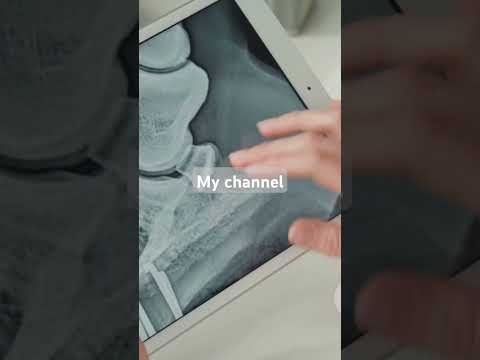
4. Radiographs (X-Rays)
Four standard views of the navicular bone are taken (lateral, DP, 65° upright pedal, and skyline/flexor views). X-rays reveal bony changes — synovial fossae, bone cysts, osteophytes, and ligament mineralization. Important caveat: many horses with radiographic navicular changes are not lame from those changes, and some horses with navicular pain have minimal radiographic changes. X-rays alone are not sufficient to diagnose navicular syndrome as the cause of lameness.
5. MRI — The Gold Standard
Standing low-field MRI (increasingly available at equine hospitals) allows visualization of the soft tissue structures that X-rays cannot show — DDFT lesions, bursal inflammation, impar ligament changes, and bone marrow edema. MRI frequently reveals that the primary pain source is a DDFT lesion or bursitis rather than the bone itself — findings that change the treatment plan. If therapeutic shoeing and medical management fail to produce improvement, MRI should be strongly considered.
6. Navicular Bursa Injection (Diagnostic / Therapeutic)
Injecting local anesthetic directly into the navicular bursa — a more specific block than the palmar digital nerve block — can confirm whether the bursa itself is the pain source. If the horse goes significantly sounder after a bursal block but not after a standard palmar digital block, bursitis is implicated. This also sets up therapeutic bursal injection as a targeted treatment option.
⚡ Quick Reference — Navicular Syndrome at a Glance
Treatment — Three Roles, Three Approaches
Navicular management is a long-term partnership between farrier and vet. The owner maintains the environment and monitoring that keeps the horse comfortable between visits.
The Farrier's Role — The Most Important Long-Term Variable
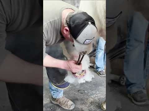
In navicular syndrome, the farrier's contribution is arguably more important than any medication or injection. The biomechanical environment of the foot — toe length, heel height, breakover position, shoeing material and style — determines how much force the navicular apparatus receives with every stride. Get the shoeing right and the horse stays comfortable. Get it wrong and no amount of medication will compensate.
The single most important trimming goal in navicular management is restoring a correct hoof-pastern axis — the hoof wall angle at the toe should be parallel to the slope of the pastern, creating a straight line from the fetlock through the hoof. In practice this means:
- Aggressively reduce toe length — the toe is the primary mechanical driver of navicular stress; shortening it reduces DDFT tension across the navicular bone with every step
- Raise the heel where appropriate — in horses with crushed, low heels, raising heel height reduces DDFT tension; however, heels that are already high should not be raised further
- Establish correct breakover — the point where the foot tips forward off the ground should be directly under the tip of the coffin bone, reducing the lever arm the toe exerts on the navicular
The egg bar shoe is the workhorse of navicular management. Its continuous oval shape extends behind the heels, providing a rear support platform that:
- Prevents heel sinking into soft footing — navicular horses often toe-land to avoid heel pain; the bar prevents this compensation from overloading the toe structures
- Distributes weight more evenly across the rear of the foot, reducing peak pressure on the navicular area
- Provides proprioceptive feedback from the frog area, encouraging the horse to use its heel again as pain decreases
- Typically fitted with a slight (2–4°) heel wedge pad in early management to further reduce DDFT tension
Rolling or rockering the toe of the shoe significantly reduces the effort and force required to break over — the moment when the heel lifts and the foot pivots forward off the toe. Less breakover effort means less peak tension on the DDFT as it crosses the navicular bone at the end of each stride. This can be done as:
- Rolled toe — the toe of the shoe is forged or ground to a rounded profile, moving breakover back toward the center of the foot
- Rocker toe — a more pronounced curve through the toe and quarter of the shoe; more relief than a simple roll
- Natural Balance shoe — a commercially available shoe built with a set-back breakover point and wide web; popular for navicular management
A wedge pad placed between the shoe and the hoof raises the heel, reducing tension on the DDFT as it curves around the navicular bone. Typical therapeutic wedge angles are 2–4°. Important cautions:
- Wedging is appropriate for horses with low, crushed, or collapsed heels — it corrects a deficit
- Wedging a horse that already has a correct or high heel angle is counterproductive — it can over-stress the suspensory apparatus and digital flexors higher up the leg
- Wedge pads are typically used as a short-to-medium-term tool while the heel is built up through trimming; the goal is eventually to maintain comfort without artificial elevation
Full sole pads placed between shoe and hoof reduce concussion and protect the sole and frog from ground contact. Pour-in polyurethane packing material fills the space completely, providing conforming cushion across the entire solar surface. This combination is particularly useful for:
- Horses working on hard, rocky, or arena footing where concussion is high
- Horses with thin soles or bruised heels complicating navicular pain
- Reducing the need for medication by improving comfort through mechanical means
Navicular horses should be shod every 5–6 weeks without exception. Allowing the cycle to stretch to 8–10 weeks permits the toe to grow long, the heels to collapse forward, and the entire corrective shoeing benefit to be lost. The farrier works with the vet to evaluate radiographs every 6–12 months and adjust the shoeing strategy as the horse's condition evolves. Consistency is the foundation of successful navicular management.
Egg Bar Shoe
Continuous oval shoe extending behind the heels. Standard first-line therapeutic shoe for navicular. Typically steel or aluminum; glue-on versions available for horses with compromised wall.
Natural Balance Shoe
Commercially designed with set-back breakover and wide web. Reduces toe leverage and improves frog contact. Popular alternative to traditional egg bar for moderate navicular cases.
Wedge Pads (2–4°)
EVA or leather heel elevation pads fitted between shoe and hoof. Reduce DDFT tension in horses with low or crushed heels. Used alongside egg bar or bar shoe.
Pour-In Pad (Vettec Equi-Pak)
Polyurethane packing that conforms to the entire solar surface. Reduces concussion, protects heel region, and improves comfort on hard footing. Used with full pads or independently.
The best navicular outcomes come from a farrier and vet who communicate directly — sharing radiographs, discussing the trim strategy, and adjusting the plan together. If your farrier has never seen your horse's X-rays, or your vet has never discussed shoeing goals with your farrier, fix that at the next visit. These two professionals working independently, rather than as a team, is the most common reason navicular management fails.
The Veterinarian's Role — Diagnosis, Pain Management, Disease Modification
The vet leads diagnosis and medical management. For navicular syndrome, the goal of veterinary treatment is reducing inflammation, slowing degeneration, and managing pain — enabling the horse to use its feet correctly so that the farrier's biomechanical corrections can take effect.
Phenylbutazone (bute) is the most commonly used NSAID in navicular management — reducing inflammation in the navicular bursa and surrounding tissues, and providing pain relief that allows the horse to move more freely and load the heel correctly. Long-term daily bute is associated with gastric ulcer and kidney risks; the lowest effective dose is used, often in combination with gastric protectants. Some vets prefer flunixin or firocoxib (Equioxx) for longer-term use due to a better safety profile.
The navicular bursa and the distal interphalangeal (coffin) joint are the two primary injection targets in navicular cases:
- Navicular bursa injection — triamcinolone or methylprednisolone with hyaluronic acid; directly targets the navicular apparatus; typically provides 3–6 months of relief; repeated as needed
- Coffin joint injection — the coffin joint communicates with the navicular bursa in many horses; injecting here reaches both structures; technically simpler than direct bursal injection
- Hyaluronic acid alone — used in horses where steroid use is limited (e.g., laminitis risk); lubricates the bursa and joint without the anti-inflammatory potency of steroids
Two bisphosphonate products are licensed for use in horses with navicular syndrome:
- Tildren (tiludronate) — intravenous infusion; inhibits osteoclast activity (bone breakdown), slowing the degenerative remodeling of the navicular bone; clinical trials show significant improvement in lameness scores in navicular horses
- Osphos (clodronate) — intramuscular injection; same mechanism as Tildren but different delivery; similar efficacy data
Bisphosphonates are disease-modifying, not just pain-relieving — they slow the underlying degenerative process. They are not a cure, but they represent a meaningful advance in navicular treatment. Should not be used in growing horses or repeatedly at short intervals.
Isoxsuprine is a vasodilator historically used to improve blood flow to the navicular bone — based on the theory that reduced circulation contributes to navicular degeneration. The evidence base is weak by modern standards, and many equine vets have moved away from it in favor of bisphosphonates and injections. However, it remains inexpensive, has a low side-effect profile, and some horses appear to respond to it — so it continues to be used, particularly as an adjunct therapy or in horses where other treatments are cost-prohibitive.
For horses with navicular syndrome driven primarily by DDFT lesions or bursitis — particularly where MRI has identified specific soft tissue pathology — regenerative therapies are increasingly used:
- PRP (Platelet-Rich Plasma) — concentrated platelets injected into the navicular bursa or directly into a DDFT lesion; promotes healing of soft tissue damage
- IRAP (Interleukin-1 Receptor Antagonist Protein) — anti-inflammatory protein harvested from the horse's own blood; reduces joint and bursal inflammation
- Stem cell therapy — mesenchymal stem cells injected into DDFT lesions to promote regenerative repair; emerging evidence, variable results
When all conservative treatments have failed, palmar digital neurectomy — surgically cutting the palmar digital nerves to permanently desensitize the caudal heel — remains a salvage option. The horse becomes sound because it no longer feels the pain, not because the disease is treated. Important considerations:
- Neurectomy does not stop disease progression — the horse remains comfortable but the degeneration continues
- Risk of DDFT rupture in desensitized horses — the horse can no longer feel warning pain before a tendon failure; this is a serious and sometimes catastrophic complication
- The horse must be identified as neurectomized when sold; in most disciplines, competing on a neurectomized horse requires disclosure and may be prohibited
- In appropriate candidates — older horses with severe disease who simply need comfort for light activity — neurectomy can provide years of quality of life
Tildren (Tiludronate)
IV bisphosphonate licensed for navicular syndrome. Slows degenerative bone remodeling. Administered as a single slow IV infusion; effects last 6–12 months. One of the most evidence-backed navicular treatments available.
Osphos (Clodronate)
IM bisphosphonate. Same mechanism as Tildren, different delivery route. May be more practical in field settings. Similar efficacy to Tildren in clinical trials.
Corticosteroid + HA Bursal Injection
Triamcinolone or methylprednisolone with hyaluronic acid injected into the navicular bursa or coffin joint. First-line injectable treatment. Typically provides 3–6 months of significant relief.
Firocoxib (Equioxx)
COX-2 selective NSAID. Safer for long-term daily use than phenylbutazone — reduced gastric and renal risk. Appropriate for horses needing ongoing pain management over months to years.
The Horse Owner's Role — Footing, Workload, Monitoring, Consistency
Navicular syndrome is a chronic condition — there is no cure, only management. The owner's role is maintaining the environment, monitoring daily comfort, enforcing the shoeing schedule, and making intelligent workload decisions that keep the horse sound long-term rather than pushing for performance at the cost of soundness.
Footing is the single biggest environmental variable the owner controls. Hard, unyielding surfaces dramatically increase navicular loading on every stride. Practical steps:
- Arena footing — keep arena sand properly maintained at 3–4 inches depth; packed hard sand is nearly as bad as asphalt for navicular horses
- Paddock footing — add pea gravel or road base to high-traffic areas; navicular horses standing on packed dirt or gravel all day accumulate significant cumulative concussion even when not being worked
- Avoid asphalt and concrete — no schooling or hacking on hard roads without hoof protection; full pads or hoof boots for any road work
- Stall bedding — deep, supportive bedding allows the horse to rest its feet on a giving surface during stall time; don't underestimate the cumulative benefit
Navicular horses can continue working — often for years — if workload is managed sensibly:
- Warm up longer on softer footing before any demanding work
- Avoid repetitive tight circles on hard ground — this concentrates force on the inside limb's caudal heel
- Rest after injection treatments — the horse needs 2–4 weeks of reduced work after bursal injections for full effect; do not resume heavy training immediately
- Monitor the horse's response to each ride — consistent post-exercise stiffness or worsening lameness over days indicates the current workload is exceeding what the horse can manage
- Have honest conversations with your trainer and vet about realistic performance expectations; protecting soundness long-term is always worth a short-term workload reduction
A navicular horse managed on a 5–6 week shoeing cycle can look like a different horse than one allowed to go 10–12 weeks. The corrective trim and therapeutic shoe are the foundation of every other treatment. Stretching the schedule to save money or because the horse "seems okay" is a false economy — the toe grows long, the heel collapses, the navicular loading worsens, and the next veterinary visit will cost more than the saved shoeing appointment. Set a reminder and treat the shoeing schedule as non-negotiable.
Because navicular is a chronic fluctuating condition, keeping a simple daily log is genuinely useful. Note:
- Willingness to walk out from the stall in the morning — navicular horses often show morning stiffness that improves with movement
- Landing pattern — is the horse landing heel-first (good), flat (acceptable), or consistently toe-first (concerning)?
- Digital pulse — bounding pulse indicates increased inflammation in the heel region
- Post-exercise comfort vs. the previous session
Strong, well-keratinized hoof horn holds therapeutic shoes more securely and provides a better biomechanical platform. Navicular horses often have compromised hoof horn quality from years of abnormal loading. Support with:
- Biotin 20mg/day — most evidence-supported hoof supplement
- Methionine and zinc — essential for keratin cross-linking and dense horn production
- Omega-3 fatty acids — reduce systemic inflammation; some evidence for improved laminar health
- Avoid supplements marketed specifically for "navicular" without evidence — most are biotin-based rebrands at premium prices
Navicular horses given NSAIDs to control pain can feel deceptively comfortable and be pushed beyond what their damaged structures can safely tolerate. A horse that is comfortable only because of bute or injections should have its workload assessed honestly — riding a medicated horse as if it were sound can accelerate the very structural damage you are trying to manage. Use medication to support healing and correct biomechanics, not to enable performance that the horse cannot sustain without drugs.
Prevention & Slowing Progression
Navicular syndrome cannot always be prevented — genetics and conformation play a real role. But its onset can often be delayed, and its progression slowed, with consistent management.
Strict Trimming Schedule
The most effective prevention is maintaining correct hoof-pastern axis through regular trimming. Never allow the toe to grow long. For high-risk horses (Quarter Horses in performance work, horses with small feet), 5–6 week farrier visits from the start of training.
Good Footing Always
Years of work on hard, unforgiving surfaces accumulate damage to the navicular apparatus. Maintain arena footing properly. Avoid unnecessary road work without pads. This is the most controllable environmental factor in navicular prevention.
Act on Early Signs
A horse that begins toe-landing, stumbling occasionally, or showing morning stiffness in the fronts deserves a lameness evaluation — not "wait and see." Navicular diagnosed at the early soft-tissue phase responds far better to treatment than disease caught after years of bone remodeling.
Maintain Healthy Weight
Every extra kilogram of body weight increases navicular loading on every stride. An overweight performance horse is at substantially higher risk of navicular deterioration. Body condition score 4–5 of 9 is ideal for navicular-prone horses.
Pre-Purchase Evaluation
Any performance horse purchase should include a full lameness evaluation including flexion tests, nerve blocks if indicated, and radiographs of all four feet. Navicular changes on pre-purchase X-rays must be interpreted in context — some horses with radiographic changes perform for years; others are a liability. Get expert interpretation.
Hoof Quality Nutrition
Dense, quality hoof horn holds shoes better, provides better biomechanical cushion, and resists the white line disease that can complicate navicular cases. Start biotin and a balanced mineral supplement before problems appear — preventive supplementation is cheaper than remedial care.
Navicular Syndrome Videos — Watch & Learn
Farrier and veterinary perspectives on diagnosis, therapeutic shoeing, and long-term management of navicular syndrome.